Article

Formation of a Colloidal CdSe and ZnSe Quantum Dots via a Gamma Radiolytic Technique

1
Department of Physics, University Putra Malaysia, Serdang 43400, Selangor, Malaysia
2
Young Researchers and Elite Club, Shahr-e-Qods Branch, Islamic Azad University, Tehran 37541-374, Iran
3
School of Applied Physics, Faculty of Science and Technology, University Kebangsaan Malaysia, Bangi 43600, Selangor, Malaysia
*
Author to whom correspondence should be addressed.
Appl. Sci. 2016, 6(10), 278; https://doi.org/10.3390/app6100278
Submission received: 28 July 2016 / Revised: 18 September 2016 / Accepted: 23 September 2016 / Published: 28 September 2016

Abstract

:
Colloidal cadmium selenide (CdSe) and zinc selenide (ZnSe) quantum dots with a hexagonal structure were synthesized by irradiating an aqueous solution containing metal precursors, poly (vinyl pyrrolidone), isopropyl alcohol, and organic solvents with 1.25-MeV gamma rays at a dose of 120 kGy. The radiolytic processes occurring in water result in the nucleation of particles, which leads to the growth of the quantum dots. The physical properties of the CdSe and ZnSe nanoparticles were measured by various characterization techniques. X-ray diffraction (XRD) was used to confirm the nanocrystalline structure, energy-dispersive X-ray spectroscopy (EDX) was used to estimate the material composition of the samples, transmission electron microscopy (TEM) was used to determine the morphologies and average particle size distribution, and UV-visible spectroscopy was used to measure the optical absorption spectra, from which the band gap of the CdSe and ZnSe nanoparticles could be deduced.

1. Introduction

Over the past two decades, semiconductor nanoparticles or quantum dots with sizes between 1 and 20 nm, have received much attention because of the quantum confinement effect of their surface electrons. In this dimension, the particles are in the transition between the bulk and molecular regimes and contain hundreds to a few thousand atoms only. The electronic, optical, and magnetic properties of quantum dots can be tailored by changing the material composition and varying the physical size [1,2,3]. In particular, group II–VI compounds such as narrow band gap CdSe quantum dots and wider band gap ZnSe quantum dots, have become intriguing materials prospects for the development of electro-optical, optical, and biomedical devices, such as light-emitting diodes [4], photo detectors [5], lasers [6], solar cells [7], and biomedical imaging applications [8,9].
The method of synthesis and, to some extent, the local environment can influence the particle size and physical properties of quantum dots. An organic polymer containing hydrophilic functional groups, such as poly(vinyl pyrrolidone) (PVP) or poly(vinyl alcohol) (PVA), is often used as a capping agent to prevent uncontrolled growth and aggregation of the particles [10]. The particles are dispersed and suspended homogeneously in the colloidal solution [11]. Some of the techniques associated with the synthesis of colloidal CdSe and ZnSe quantum dots are chemical synthesis [12,13], photochemical synthesis [14], micro emulsion [15], low temperature methods [16], and sol-gel methods [17], among others. Most of these methods have successfully produced particles with desired sizes or shapes and electronic or optical properties. Colloidal CdSe nanoparticles synthesized using the micro emulsion method have shown narrow particle sizes between 2.10 and 2.78 nm with band gaps of 2.8 and 1.8 eV, respectively [18]. Particles in the size range of 2.8–3.0 nm with a band gap range of 2.76–2.95 eV have been prepared by the chemical method [19]. The microwave radiation method can produce larger particles in the size range of 10 to 19 nm with a narrow band gap of approximately 1.79 eV [20]. CdSe nanoparticles with sizes of 43 nm and a narrow band gap of 1.75 eV were prepared by the hydrothermal method [21]. However, the narrow size ZnSe nanoparticles of approximately 4 nm with a band gap of 3.3 eV were fabricated using the microwave method [20]. ZnSe nanoparticles of 5.14 nm with a band gap of 3.2 eV have been prepared using the micro emulsion method [22]. Larger sized particles at 11.79 nm can be prepared by chemical methods with a band gap of 3.3 eV [23]. ZnSe nanoparticles with a size between 14.3 and 17.9 nm, and with a wider band gap up to 4.48 eV, have been synthesized using hydrothermal methods [24].
The gamma radiolytic method is another technique that can be used to produce colloidal nanomaterials [25,26,27,28,29,30,31,32]. This technique was originally used for the synthesis of colloidal silver nanoparticles [25,26], colloidal platinum nanoparticles [27], and colloidal nickel-aluminum and copper-aluminum alloy nanoparticles [28,29], where the particle size of metal nanoparticles was found to decrease with increasing gamma radiation dose. This observation was explained by the competition between the nucleation and aggregation processes, where higher-dose nucleation dominates over aggregation, which allows for particle growth in which smaller particles are likely to be produce [30]. Recently, colloidal cadmium sulfide (CdS) and zinc sulfide (ZnS) quantum dots in aqueous PVA solution were fabricated using the same method [31,32]. The particle size of colloidal CdS nanoparticles was found to increase from 13.31 nm at 10 kGy to 21.07 nm at 40 kGy with band gaps of 3.15 and 3.03 eV respectively [31].The particle size of the colloidal ZnS nanoparticles was also found to increase from 53 to 59 nm and the band gap decreased from 3.74 to 3.68 eV upon increasing the dose from 10 to 50 kGy [32]. The particle sizes of colloidal CdS and ZnS quantum dots appeared to increase with increasing dose, indicating that at higher doses the aggregation process is favored over the nucleation process, producing larger particle sizes.
In light of the above results, this report is aimed at creating colloidal CdSe and ZnSe quantum dots via a gamma radiolytic technique. The motivation is that the method is simple and efficient at producing quantum dots. The nucleation and aggregation processes can occur during the irradiation time, normally lasting for several hours or even days depending on the dose and source strength. The formation of quantum dots is immediately halted when the source is removed, resulting in very good particle stability for further particle characterization over a several-day period [25,26,27,28,29,30,31,32].

2. Materials and Methods

The starting materials for the preparation of CdSe and ZnSe nanoparticles were cadmium sulfite (CdSO3·8H2O), Zinc sulfite (ZnSO3·8H2O), selenium powder (99.999%), ethylenediamine, isopropyl alcohol (IPA) and polyvinyl pyrrolidone (PVP Mw = 10,000). Deionized water was also used as a solvent. PVP was chosenas the polymer of choice in this work because of its good processability, flexibility and high transmittance [10]. All of the materials mentioned above were purchased from Sigma Aldrich (Darmstadt, Germany).
A single-step radiolytic technique based on 60Co gamma irradiation was applied in the preparation of CdSe and ZnSe nanoparticles. The first step was the dissolution of 2 g of PVP in 70 mL of deionized water, which was constantly stirred for 120 min at 70 °C. The PVP solution was then enhanced with 0.4 mmol of cadmium/zinc sulfite with simultaneous stirring, and the obtained solution was subjected to homogenization for half an hour. The subsequent step was the dissolution of 0.4 mmol of selenium powder in 80 mL of ethylenediamine. The polymer solution was then enhanced with the selenium solution and 2 mL of IPA, with continuous stirring until a homogeneous solution was obtained. Prior to irradiation at a dose of 120 kGy, oxygen was removed from the solution through nitrogen gas bubbling for 3 h.

3. Characterization

XRD patterns were recorded on a Shimadzu X-ray diffractometer XRD-3A with Cu-Kα radiation (Shimadzu model 6000, Lelyweg1, Almelo, The Netherlands), λ = 0.15418 nm. Energy-dispersive X-ray (EDX) spectroscopy was performed using an EDX spectrometer (7353, Oxford Instruments, Oxfordshire, UK). TEM was performed using a JEOL-JEM 200CX instrument (JEOL TEM model 2010F UHR, Munich, Germany) and optical absorption spectra were recorded using a UV-Visible spectrometer Shimadzu-UV1650PC (Shimadzu model UV-3600, Kyoto, Japan) in the wavelength range of 250 to 400 nm.

4. Results and Discussion

4.1. Formation Mechanism of CdSe and ZnSe Nanoparticles

In general, 1.25-MeV 60Co gamma rays interact with matter predominantly by absorption events (photoelectric absorption and pair production) and scattering events (coherent Rayleigh scattering and incoherent Compton scattering). These interactions result in the formation of secondary electrons in aqueous solution, mainly originating from Compton scattering, which consequently induce among others, hydrated electrons ( e e q ), hydroxyl radicals ( OH * ), and hydrogen radicals ( H * ) by radiolysis of water according to [25,26,27,28,29,30,31,32]:
H 2 O     e e q ,   H + ,   H 3 O ,   H * ,  OH * ,   H 2 ,   H 2 O 2       ( Radiolysis of water )
The hydrated electrons, e aq , are strong reducing agents, and reduce selenium into selenide (Se2−):
Se0 + 2eaq → Se2−
In the aqueous solution, cadmium sulfite or zinc sulfite can dissociate into positive Cd2+ or Zn2+ cations and negative SO32− anions:
CdSO3 → Cd2+ + SO32− or ZnSO3 → Zn2+ + SO32−
The Se2− ions can be reacted with Cd2+ or Zn2+ ions to generate CdSe or ZnSe nanoparticles throughout the reactant medium via the nucleation process:
Cd2+ + Se2− → CdSe0 or Zn2+ + Se2− → ZnSe0
Individual CdSe or ZnSe compounds begin to grow in the following aggregation process:
CdSe0 + CdSe0 → CdSe02 or ZnSe0 + ZnSe0 → ZnSe02
CdSe0 + CdSe0n → CdSe0n+1 or ZnSe0 + ZnSe0m → ZnSe0m+1
where n and m can be hundreds to a few thousand depending on the particle size.
Hydroxyl and hydrogen radicals ( OH * and H * ) are also strong oxidizing agents in aqueous colloidal solution. Therefore, to prevent their reactions, isopropanol (IPA) was added to the precursor solutions [27]. All organics in the reactant medium including PVP and IPA, can scavenge OH * and H * radicals and can simultaneously change into PVP and IPA radicals by the following reaction [33].
PVP + OH* → PVP* + H2O; PVP* + H* → PVP* + H2; IPA* + PVP* → crosslinked PVP gel
IPA + OH* → IPA* + H2O; IPA* + H* → IPA* + H2; IPA* + IPA* → crosslinked IPA gel

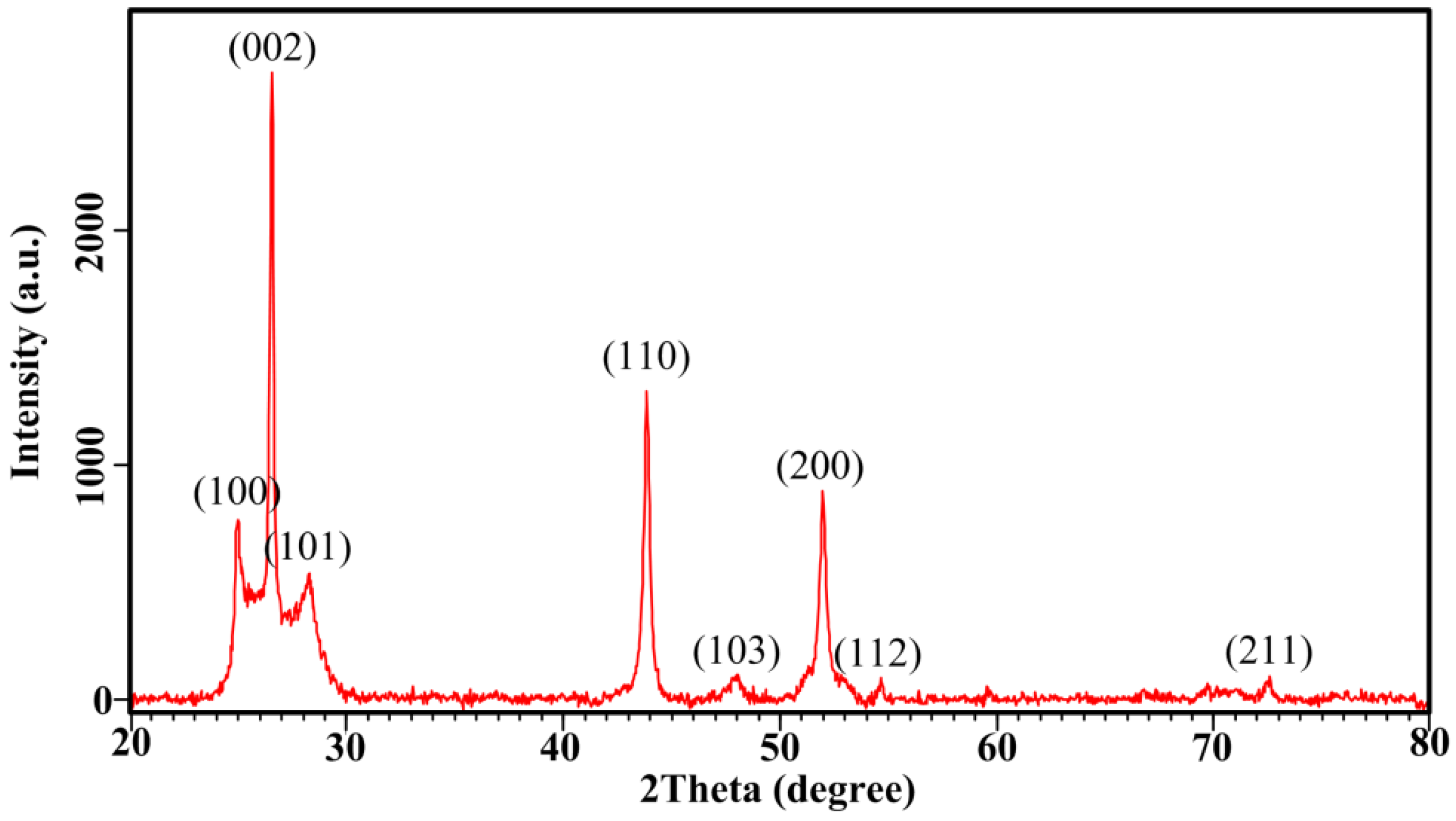
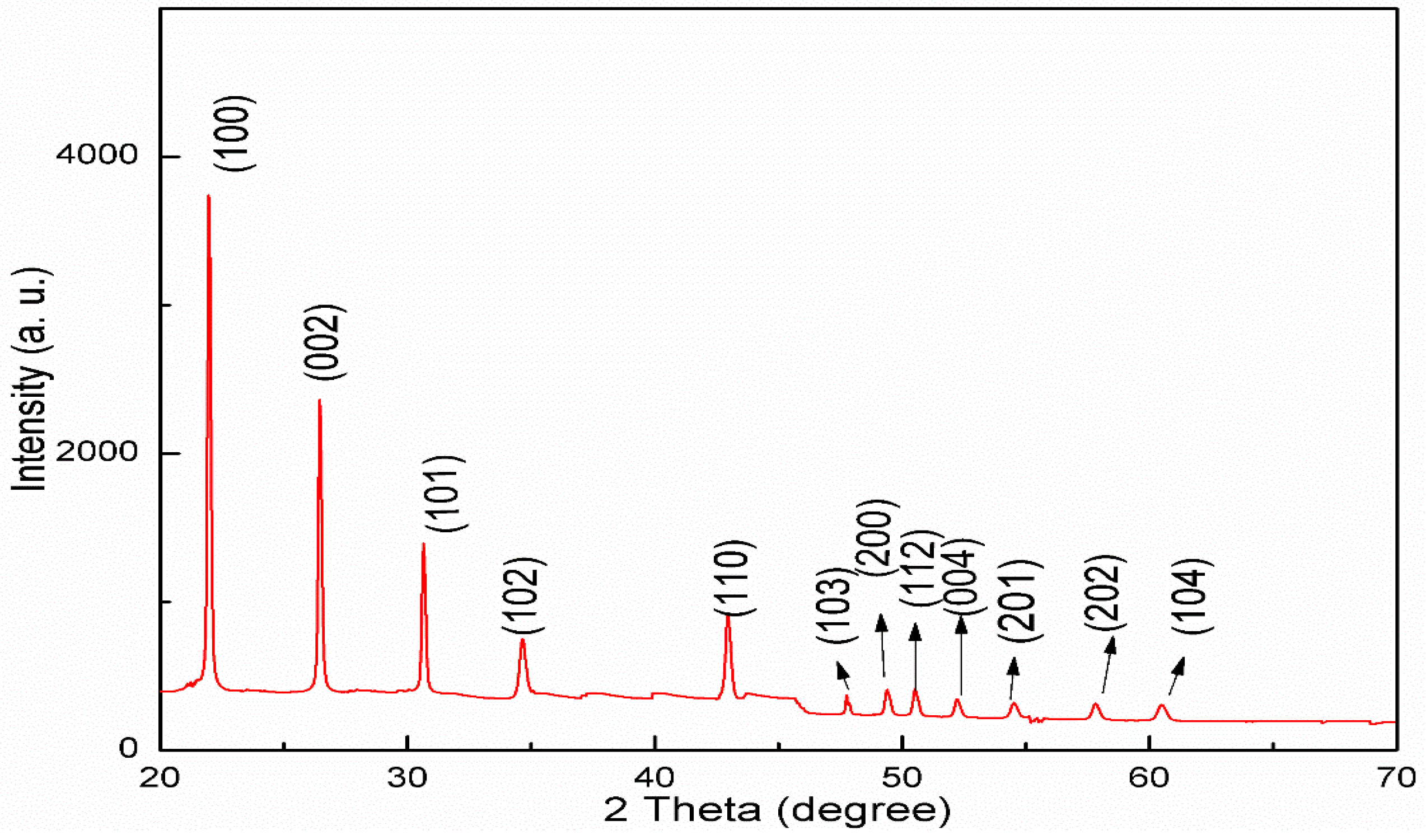
4.2. Structural Analysis (XRD)

The crystal structure and crystallite size of the synthesized nanoparticles were investigated by generating diffraction patterns, XRD, from the crystalline powder samples at ambient temperature.
Figure 1 shows the XRD pattern of CdSe nanoparticles synthesized with a 120 kGy dose showing the formation of the nanoparticles. The peaks observed in the XRD patterns at 2θ value of 25.1, 26.5, 28.2, 36.8, 43.8, 47.9, 51.9, 54.6 and 72.4° nearly match with the (100), (002), (101), (110), (103), (200), (112) and (211) crystalline planes, respectively, of the hexagonal CdSe structure reported in The International Centre for Diffraction Data (ICDD) PDF 02-0330.
From the width of the XRD peak, the mean crystalline size can be calculated using the Debye-Scherrer equation [34]:
D = kλ/βcosθ
where k is a constant equal to 0.9, λ is the wavelength of the X-ray radiation (0.1542 nm), θ is the diffraction angle, and β is the full width half maximum (FWHM). The average crystalline size of CdSe nanoparticles was determined from the full width half maximum of the (002) reflection of the XRD pattern using the Debye-Scherrer equation. The calculation showed that the average crystalline size is 16.3 nm. Figure 2 shows the XRD pattern of ZnSe nanoparticles synthesized with a 120 kGy dose. The strong and sharp peaks at 2θ values of 26.40, 28.60, and 30.50° virtually match those of the (100), (002) and (101) crystalline planes, respectively, of the hexagonal ZnSe structure reported in the ICDD PDF 89-2940. Furthermore the peaks observed at 2θ values of 34.9, 43.6, 47.2 49.3, 50.5, 51.9, 54.4, 57.7 and 60.4° were also assigned to the (102), (110), (103), (200), (112), (201), (004), (202) and (104) crystalline planes of the hexagonal phase ZnSe, respectively. However, some extra peaks not identified as ZnSe may be attributed to some impurity such as a zinc complex formed during the reactions.
The average crystallite size of the ZnSe nanoparticles was calculated to be 10.7 nm from the sharp reflection peaks of (100) using the Debye-Scherrer equation.

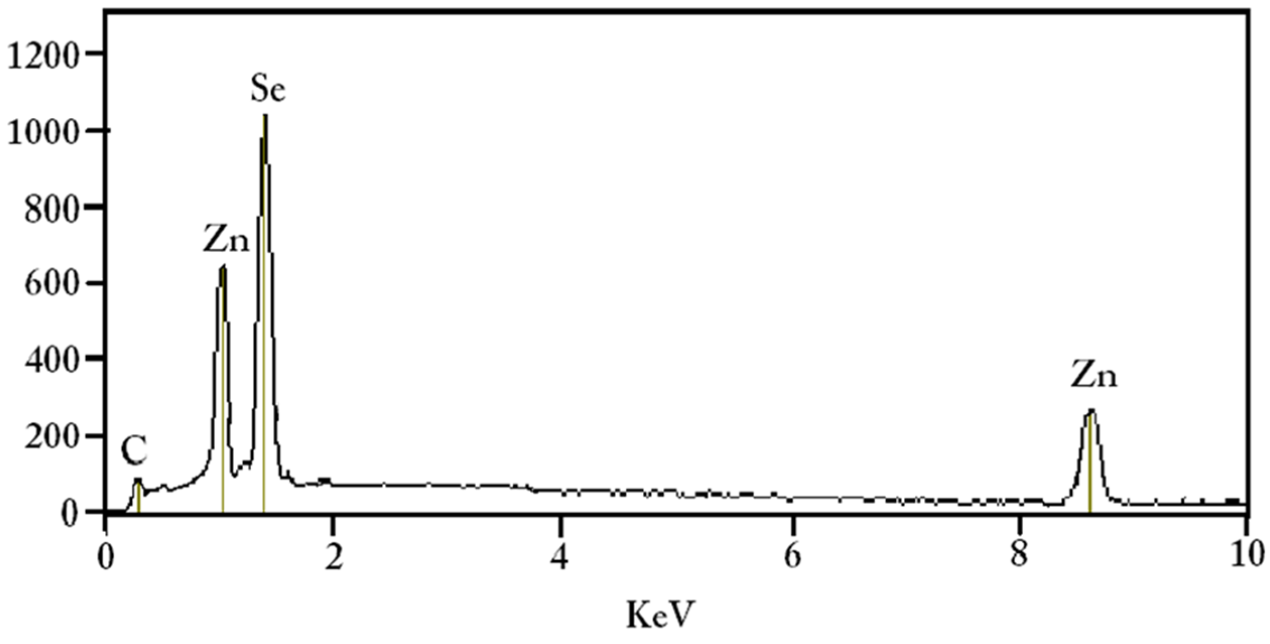
4.3. Elemental Composition Analysis (EDX)

Energy dispersive X-ray (EDX) analysis was performed to confirm the quantitative composition of elements in the CdSe and ZnSe nanoparticles after irradiation with 120 kGy. The EDX patterns are shown in Figure 3 and Figure 4 and the related data are listed in Table 1. The host material of CdSe nanoparticles exhibits four elemental peaks; three for cadmium located at ~0.4, 3.1 and 3.3 keV, and one for Se located at 1.4 keV. For ZnSe nanoparticles, two elemental peaks related to Zn were found at ~1.0 and 8.6 keV, and one for Se at ~1.4 keV. The existence of a low intensity peak at ~0.3 keV in both spectra is attributed to the carbon film used as a holder for the samples during EDX measurements. Data analysis from EDX patterns (Table 1) confirmed the stoichiometric composition of elements in the samples. One of the advantages of nanomaterials synthesized by the gamma radiolytic method is that the particles produced are pure, as indicated by their elemental composition, which has been established in previous works [25,26,27,28,29,30,31,32].

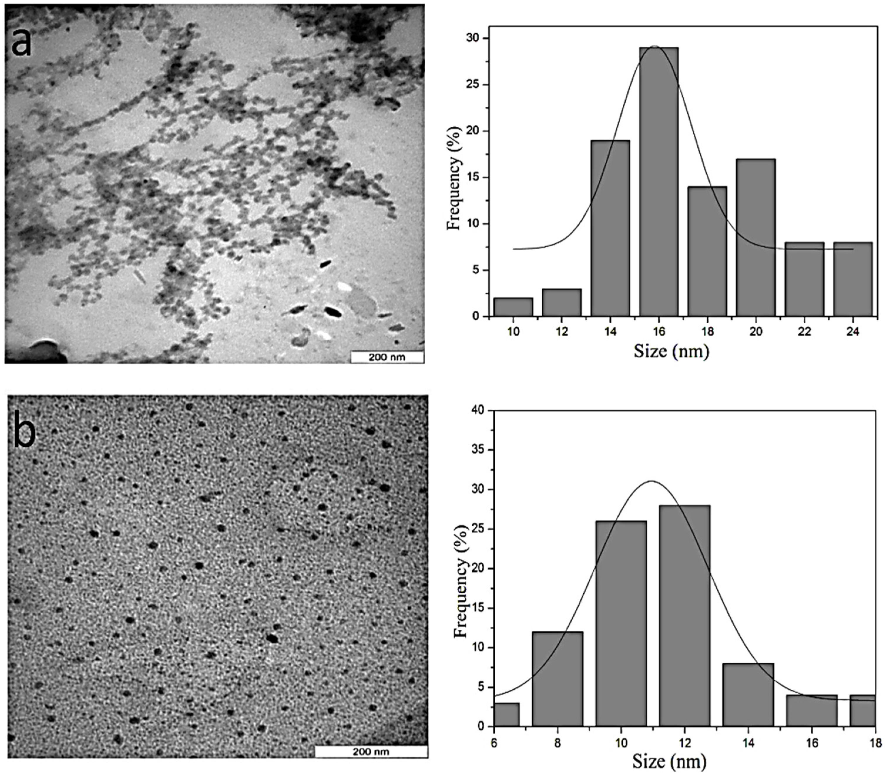
4.4. Morphology and Particle Size

Figure 5 shows the TEM images and corresponding size distribution histogram of CdSe and ZnSe nanoparticles. From the illustration, a uniform spherical morphology with a relatively narrow homogeneous distribution was achieved for both the CdSe and ZnSe nanoparticles. The average particle size of the CdSe nanoparticles was estimated to be 17.3 nm whereas the average particle size of the ZnSe nanoparticles was found to be 11.2 nm. The average particle size obtained from TEM images are in good agreement with the estimated crystallite size from the XRD profile results for both CdSe and ZnSe nanoparticles (Table 2). At 120 kGy, the gamma radiolytic method produced CdSe and ZnSe nanoparticles with larger particle sizes compared to those synthesized by microwave [20] or hydrothermal [21,24] methods. The creation of hydrated electrons by gamma irradiation in the colloidal solution is considered constant for formation of both CdSe and ZnSe nanoparticles, but the particle size of CdSe nanoparticles is greater than that of ZnSe nanoparticles. The fundamental reason for this difference is that the atomic size of Cd (atomic mass of 112.40) is greater than that of the atomic size of Zn (atomic mass of 55.87). Another advantage of the gamma radiolytic method is that the formation of CdSe and ZnSe nanoparticles ceases immediately after removal of the gamma ray source. The formed nanoparticles remain stable (in our case for 7–10 days) prior to UV-Visible absorption and TEM measurements. Similar findings have been reported in previous investigations [31,32].
The ZnSe and CdSe nanoparticles were found to increase in size with increasing dose [31,32]. However, increasing the molecular mass or concentration of PVP reduced the particle size of ZnSe and CdSe nanoparticles as a result of the efficient capping mechanism making the individual particles difficult to aggregate [10]. An increase in precursor concentration would increase the number of nucleation processes of ZnSe and CdSe nanoparticles because of the increasing number of Cd, Zn, and Se ions, thus enlarging the particle size [28].

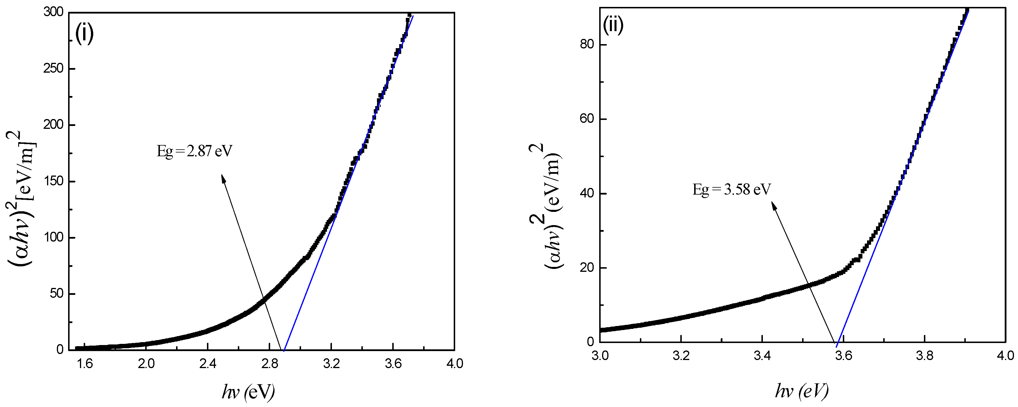
4.5. Optical Properties

The optical band gap energy of the CdSe and ZnSe nanoparticles was estimated through the Tauc equation [35]:
( α h v ) 1 / n = B ( h v E g )
where α is the absorption coefficient, is the photon energy of the incident light, Eg is the band gap energy, B is a constant and n = 1/2, 1, 3/2 or 2 for allowed direct, allowed indirect, forbidden direct and forbidden indirect, respectively. The absorption band gap of prepared samples was estimated by extrapolating the linear portion of (α)2 as a function of hν. Then, the intercept of the extrapolated line on the x-axis indicates the band gap energy of the sample. The values of (αhv)2 versus (hv) were plotted using the UV-Visible spectra of the samples as illustrated in Figure 6. Straight lines were drawn to fit the experimental band gap curves and were extended intersect the (hv) axis to determine the optical band gap values of the CdSe and ZnSe nanoparticles.
Careful observation of Figure 6 shows that the band gap energy for the CdSe and ZnSe nanoparticles can be extrapolated to 2.87 eV and 3.58 eV respectively. These results shows that the magnitudes of the obtained band gap energies for both CdSe and ZnSe nanoparticles are higher than the band gap energies of their bulk counterparts of 1.74 and 2.7 eV, respectively—the blue shift phenomenon.

5. Conclusions

Colloidal CdSe and ZnSe quantum dots were synthesized using the radiolytic technique by gamma irradiation in which the hydrated electrons that are generated in water are responsible for the formation of CdSe and ZnSe nanoparticles. EDX was used to confirm the stoichiometric elemental composition of Zn, Cd and Se in the samples. The X-ray powder diffraction patterns reveal the hexagonal crystal structure of both CdSe and ZnSe nanoparticles and the average crystallite sizes were calculated to be 16.3 nm and 10.7 nm, respectively, from Scherrer’s equation. The TEM micrographs show that CdSe and ZnSe nanoparticles are spherical in shape with average diameter of 17.3 and 11.2 nm, respectively. The optical band gaps of the samples, which were determined from UV-Visible absorption spectroscopy were found to be 2.87 and 3.58 eV for the CdSe and ZnSe nanoparticles, respectively.

Acknowledgments

The authors extend their sincere gratitude to University Putra Malaysia for providing financial support and facilities for the completion of this work.

Author Contributions

Aeshah Salem, Elias Saion and Naif Mohammed Al-Hada conceived and designed the experiments; Aeshah Salem performed the experiments; Aeshah Salem, Elias Saion, Naif Mohammed Al-Hada and Nayereh Soltani analyzed the data; Aeshah Salem, Elias Saion and Naif Mohammed Al-Hada contributed reagents/materials/analysis tools; Aeshah Salem, Elias Saion, Naif Mohammed Al-Hada, Abdul Halim Shaari, Halimah Mohamed Kamari, Nayereh Soltani and Shahidan Radiman wrote the paper.

Conflicts of Interest

The authors declare no conflict of interest.

References

  1. Talapin, D.V.; Lee, J.-S.; Kovalenko, M.V.; Shevchenko, E.V. Prospects of colloidal nanocrystals for electronic and optoelectronic applications. Chem. Rev. 2009, 110, 389–458. [Google Scholar] [CrossRef] [PubMed]
  2. Suresh, S. Semiconductor nanomaterials, methods and applications: A review. Nanosci. Nanotechnol. 2013, 3, 62–74. [Google Scholar]
  3. Boles, M.A.; Ling, D.; Hyeon, T.; Talapin, D.V. The surface science of nanocrystals. Nat. Mater. 2016, 15, 141–153. [Google Scholar] [CrossRef] [PubMed]
  4. Chen, H.-S.; Hsu, C.-K.; Hong, H.-Y. Ingan-cdse-znse quantum dots white LEDs. IEEE Photonics Technol. Lett. 2006, 18, 193–195. [Google Scholar] [CrossRef]
  5. Žídek, K.; Abdellah, M.; Zheng, K.; Pullerits, T. Electron relaxation in the CdSe quantum dot-ZnO composite: Prospects for photovoltaic applications. Sci. Rep. 2014, 4. [Google Scholar] [CrossRef] [PubMed]
  6. Gronin, S.; Zverev, M.; Sorokin, S.; Sedova, I.; Gamov, N.; Peregoudov, D.; Studionov, V.; Kop’ev, P.; Ivanov, S. Effective green semiconductor lasers with multiple CdSe/ZnSe QD active region for electron beam pumping. Acta Phys. Pol. A 2008, 114, 1115–1122. [Google Scholar] [CrossRef]
  7. Saidov, A.; Amonov, K.; Kutlimurotov, B. Direct solar conversion to electricity nanoscale effects in pSi–n(Si2)1–x(ZnSe)x (0 ≤ x ≤ 0.01) of solar cells. Appl. Sol. Energy 2016, 52, 1–4. [Google Scholar] [CrossRef]
  8. Jiang, F.; Muscat, A.J. Ligand-controlled growth of ZnSe quantum dots in water during ostwald ripening. Langmuir 2012, 28, 12931–12940. [Google Scholar] [CrossRef] [PubMed]
  9. William, W.Y.; Chang, E.; Drezek, R.; Colvin, V.L. Water-soluble quantum dots for biomedical applications. Biochem. Biophys. Res. Commun. 2006, 348, 781–786. [Google Scholar]
  10. Soltani, N.; Saion, E.; Erfani, M.; Rezaee, K.; Bahmanrokh, G.; Drummen, G.P.; Bahrami, A.; Hussein, M.Z. Influence of the polyvinyl pyrrolidone concentration on particle size and dispersion of ZnS nanoparticles synthesized by microwave irradiation. Int. J. Mol. Sci. 2012, 13, 12412–12427. [Google Scholar] [CrossRef] [PubMed]
  11. Steigerwald, M.L.; Alivisatos, A.P.; Gibson, J.; Harris, T.; Kortan, R.; Muller, A.; Thayer, A.; Duncan, T.; Douglass, D.; Brus, L.E. Surface derivatization and isolation of semiconductor cluster molecules. J. Am. Chem. Soc. 1988, 110, 3046–3050. [Google Scholar] [CrossRef]
  12. Chen, H.-S.; Lo, B.; Hwang, J.-Y.; Chang, G.-Y.; Chen, C.-M.; Tasi, S.-J.; Wang, S.-J.J. Colloidal znse, ZnSe/ZnS, and ZnSe/ZnSeS quantum dots synthesized from zno. J. Phys. Chem. B 2004, 108, 17119–17123. [Google Scholar] [CrossRef]
  13. Aboulaich, A.; Geszke, M.; Balan, L.; Ghanbaja, J.; Medjahdi, G.; Schneider, R. Water-based route to colloidal mn-doped ZnSe and core/shell ZnSe/ZnS quantum dots. Inorg. Chem. 2010, 49, 10940–10948. [Google Scholar] [CrossRef] [PubMed]
  14. Zhu, J.; Liao, X.; Zhao, X.; Wang, J. Photochemical synthesis and characterization of CdSe nanoparticles. Mater. Lett. 2001, 47, 339–343. [Google Scholar] [CrossRef]
  15. Entezari, M.H.; Ghows, N. Micro-emulsion under ultrasound facilitates the fast synthesis of quantum dots of cds at low temperature. Ultrason. Sonochem. 2011, 18, 127–134. [Google Scholar] [CrossRef] [PubMed]
  16. Senthilkumar, K.; Kalaivani, T.; Kanagesan, S.; Balasubramanin, V. Low temperature method for synthesis of starch-capped ZnSe nanoparticles and its characterization studies. J. Appl. Phys. 2012, 112. [Google Scholar] [CrossRef]
  17. Gaeeni, M.R.; Tohidian, M.; Majles-Ara, M. Green synthesis of CdSe colloidal nanocrystals with strong green emission by the sol–gel method. Ind. Eng. Chem. Res. 2014, 53, 7598–7603. [Google Scholar] [CrossRef]
  18. Guleria, A.; Singh, S.; Rath, M.; Singh, A.; Adhikari, S.; Sarkar, S. Tuning of photoluminescence in cadmium selenide nanoparticles grown in CTAB based quaternary water-in-oil microemulsions. J. Lumin. 2012, 132, 652–658. [Google Scholar] [CrossRef]
  19. Amiri, G.R.; Fatahian, S.; Mahmoudi, S. Preparation and optical properties assessment of CdSe quantum dots. Mater. Sci. Appl. 2013, 4, 134–137. [Google Scholar] [CrossRef]
  20. Klauke, K.; Hahn, B.; Schütte, K.; Barthel, J.; Janiak, C. Bis ((dialkylamino) alkylselenolato) metal complexes as precursors for microwave-assisted synthesis of semiconductor metal selenide nanoparticles of zinc and cadmium in the ionic liquid [bMIm][BF4]. Nano-Struct. Nano-Objects 2015, 1, 24–31. [Google Scholar] [CrossRef]
  21. Xi, L.; Lam, Y.M.; Xu, Y.P.; Li, L.-J. Synthesis and characterization of one-dimensional cdse by a novel reverse micelle assisted hydrothermal method. J. Colloid Interface Sci. 2008, 320, 491–500. [Google Scholar] [CrossRef] [PubMed]
  22. Yang, L.; Liu, L.; Xiao, D.; Zhu, J. Preparation and characterization of ZnSe nanocrystals by a microemulsion-mediated method. Mater. Lett. 2012, 72, 113–115. [Google Scholar] [CrossRef]
  23. Deshpande, M.; Chaki, S.; Patel, N.; Bhatt, S.; Soni, B. Study on nanoparticles of ZnSe synthesized by chemical method and their characterization. J. Nano Electron. Phys. 2011, 3, 193–202. [Google Scholar]
  24. Zeng, Q.; Xue, S.; Wu, S.; Gan, K.; Xu, L.; Han, J.; Zhou, W.; Shi, Y.; Zou, R. Synthesis, field emission and optical properties of znse nanobelts, nanorods and nanocones by hydrothermal method. Mater. Sci. Semicond. Process. 2015, 31, 189–194. [Google Scholar] [CrossRef]
  25. Naghavi, K.; Saion, E.; Rezaee, K.; Yunus, W.M.M. Influence of dose on particle size of colloidal silver nanoparticles synthesized by gamma radiation. Radiat. Phys. Chem. 2010, 79, 1203–1208. [Google Scholar] [CrossRef]
  26. Saion, E.; Gharibshahi, E.; Naghavi, K. Size-controlled and optical properties of monodispersed silver nanoparticles synthesized by the radiolytic reduction method. Int. J. Mol. Sci. 2013, 14, 7880–7896. [Google Scholar] [CrossRef] [PubMed]
  27. Gharibshahi, E.; Saion, E. Influence of dose on particle size and optical properties of colloidal platinum nanoparticles. Int. J. Mol. Sci. 2012, 13, 14723–14741. [Google Scholar] [CrossRef] [PubMed]
  28. Abedini, A.; Larki, F.; Saion, E.; Zakaria, A.; Hussein, M.Z. Influence of dose and ion concentration on formation of binary al–ni alloy nanoclusters. Radiat. Phys. Chem. 2012, 81, 1653–1658. [Google Scholar] [CrossRef]
  29. Abedini, A.; Larki, F.; Saion, E.; Noroozi, M. Effect of Cu2+/Al3+ mole ratio on structure of Cu–Al bimetallic nanoparticles prepared by radiation induced method. Kerntechnik 2013, 78, 214–219. [Google Scholar] [CrossRef]
  30. Abedini, A.; Daud, A.R.; Hamid, M.A.A.; Othman, N.K.; Saion, E. A review on radiation-induced nucleation and growth of colloidal metallic nanoparticles. Nanoscale Res. Lett. 2013, 8. [Google Scholar] [CrossRef] [PubMed]
  31. Kharazmi, A.; Saion, E.; Faraji, N.; Soltani, N.; Dehzangi, A. Optical properties of CdS/PVA nanocomposite films synthesized using the gamma-irradiation-induced method. Chin. Phys. Lett. 2013, 30, 1–5. [Google Scholar] [CrossRef]
  32. Kharazmi, A.; Faraji, N.; Hussin, R.M.; Saion, E.; Yunus, W.M.M.; Behzad, K. Structural, optical, opto-thermal and thermal properties of ZnS–PVA nanofluids synthesized through a radiolytic approach. Beilstein J. Nanotechnol. 2015, 6, 529–536. [Google Scholar] [CrossRef] [PubMed]
  33. Khampieng, T.; Brikshavana, P.; Supaphol, P. Silver nanoparticle-embedded poly (vinyl pyrrolidone) hydrogel dressing: Gamma-ray synthesis and biological evaluation. J. Biomater. Sci. Polym. Ed. 2014, 25, 826–842. [Google Scholar] [CrossRef] [PubMed]
  34. Cullity, B.D. Elements of X-ray Diffraction, 2nd ed.; Addison-Wesley: London, UK, 1978. [Google Scholar]
  35. Simmons, B.A.; Li, S.; John, V.T.; McPherson, G.L.; Bose, A.; Zhou, W.; He, J. Morphology of CdS nanocrystals synthesized in a mixed surfactant system. Nano Lett. 2002, 2, 263–268. [Google Scholar] [CrossRef]
Figure 1. X-ray diffraction pattern of CdSe nanoparticles.
Applsci 06 00278 g001
Figure 2. X-ray diffraction pattern of ZnSe nanoparticles.
Applsci 06 00278 g002
Figure 3. Energy dispersive X-ray (EDX) compositional analysis of CdSe nanoparticles.
Applsci 06 00278 g003
Figure 4. EDX compositional analysis of ZnSe nanoparticles.
Applsci 06 00278 g004
Figure 5. Transmission electron microscopy (TEM) for as prepared (a) CdSe and (b) ZnSe nanoparticles.
Applsci 06 00278 g005
Figure 6. Optical band gap energy for the as prepared (i) CdSe and (ii) ZnSe nanoparticle.
Applsci 06 00278 g006
Table 1. Weight percentages of elements in CdSe and ZnSe nanoparticles samples.
SampleElementWt/%
CdSeSe34.27
Cd50.47
C15.26
ZnSeSe46.13
Zn33.65
C20.22
Table 2. Crystal structure, size and optical band gap of CdSe and ZnSe nanoparticles.
SampleCrystal StructureCrystallite Size/nmParticle Size/nmBandgap /eV
CdSeHexagonal16.317.32.87
ZnSeHexagonal10.711.23.58

